
Cryopreservation Artifacts: How Poor Freezing Protocols Mask T-Cell Responses
- No Comments
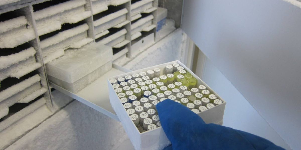
One of the most frustrating scenarios in bioanalytics is the “High Background” plate. You run a functional assay, and your negative control wells are full of non-specific activity. Is it the assay? Is it the reagents?
More often than not, it is the cryopreservation protocol. Through our PBMC Processing & Functional Validation Services at Accelevir, we frequently see that the root cause of assay failure isn’t the assay itself—it’s the stress placed on the PBMCs during the freezing process.
The Cellular Stress Artifact
When PBMCs are frozen incorrectly, cells undergo extreme membrane stress. Studies in Cytometry Part A have demonstrated that upon thawing and plating into an assay, these stressed cells can release a burst of non-specific cytokines or apoptotic bodies.
In the assay, this looks like valid data, reducing the signal-to-noise ratio and making it impossible to detect low-frequency T-cell responses.
Standardizing the Curve
Standardizing the freezing curve is non-negotiable. Whether a clinical site is using a Passive Freezing Container (Mr. Frosty) or a true Controlled-Rate Freezer, the results will vary wildly. Additionally, the choice between simple 10% DMSO + FBS versus a validated Commercial Cryomedium impacts post-thaw functional recovery.
Conclusion
You cannot fix bad cells with a good assay. To ensure your T-cell data is real, you must evaluate the cryopreservation process as rigorously as the analytical method.
Stop losing data to background noise. Start your project and provide your sample parameters to begin an independent functional validation of your biobank today.